Fdp Anatomy Hand / Jersey Finger Wikipedia - This condition is caused by excessive use of hand, resulting in pain and swelling on the dorsum of the hand.
Get link
Facebook
X
Pinterest
Email
Other Apps
Fdp Anatomy Hand / Jersey Finger Wikipedia - This condition is caused by excessive use of hand, resulting in pain and swelling on the dorsum of the hand.. The extensor indicis proprius and extensor digiti minimi lie ulnar to the extensor. The forearm consists of two bones, the radius and the ulna. Skin of the medial 1 1/2 digits This condition is caused by excessive use of hand, resulting in pain and swelling on the dorsum of the hand. J hand surg am 1989;
Within the wrist and hand, the fds tendons are located superficially (volar) to the fdp tendons. Muscles flexor digitorum profundus (fdp) functions as a flexor of the dip joint; These tendons enter the hand via the carpal tunnel, enclosed in a common synovial sheath. Originates at medial epicondyle and inserts at flexor retinaculum/palmar aponeurosis. The flexor tendons of the hand and fingers pass through the carpal tunnel of the wrist before separating in the palm and moving toward their ultimate attachment sites in the fingers.
Skeletal Anatomy Of The Hand Hand Clinics from els-jbs-prod-cdn.jbs.elsevierhealth.com (ulnar half), abductor digiti minimi m., flexor digiti minimi brevis m., opponens digiti minimi m., ulnar 2 lumbrical mm., palmar and dorsal interosseous mm. Flexor digitorum superficialis (fds) and flexor digitorum profundus (fdp). Each finger has two flexor tendons, the flexor digitorum superficialis (fds) and the flexor digitorum profundus (fdp). It is considered an extrinsic hand muscle because it acts on the hand while its muscle belly is located in the forearm. Compartment syndrome rarely occurs in the first dorsal interosseous compartment of hand. Absent in about 1/7 of population. This muscle extends from the proximal part of the ulna to the distal phalanges of the 2nd to 5th digit. The forearm consists of two bones, the radius and the ulna.
Ain i & m, ulnar r & l
(ulnar half), abductor digiti minimi m., flexor digiti minimi brevis m., opponens digiti minimi m., ulnar 2 lumbrical mm., palmar and dorsal interosseous mm. The ring finger is involved in 75% of cases. This condition is caused by excessive use of hand, resulting in pain and swelling on the dorsum of the hand. The forearm consists of two bones, the radius and the ulna. The extensor indicis proprius and extensor digiti minimi lie ulnar to the extensor. The long flexor tendons of the fingers arise from the flexor digitorum superficialis (fds) and flexor digitorum profundus (fdp) forearm muscles. The flexor digitorum profundus (fdp) is an extrinsic hand muscle that flexes the metacarpophalangeal and distal interphalangeal joints of the index, middle, ring, and little fingers. Leddy and packer classification for zone i flexor tendon injury: Learn vocabulary, terms, and more with flashcards, games, and other study tools. The treatment of zone i flexor digitorum profundus (fdp) avulsions and lacerations requires reattachment of the tendon to the distal phalanx. This complex structure connects the entire hand to the radius and ulna, facilitates the passage of tendons together with the above mentioned neurovascular structures from the forearm to the hand, and permits us to exploit all its movements. Originates at medial epicondyle and inserts at flexor retinaculum/palmar aponeurosis. Hand and finger ultrasound education showing how to, scanning protocol, normal anatomy, anatomic variants, tendon, pulley googhywoiu9839t543j0s7543uw1.
Flexor carpi ulnaris m., flexor digitorum profundus m. Within the fingers, the fds tendons split at the level of the mid diaphysis of the proximal phalanges to allow the fdp tendons to pass superficially. It is considered an extrinsic hand muscle because it acts on the hand while its muscle belly is located in the forearm. Flexor digitorum superficial splits into radial and ulnar slips prior to insertion on middle phalanx. It is considered an extrinsic hand muscle because it acts on the hand while its muscle belly is located in the forearm.
Jersey Finger Blog Nuem Blog from images.squarespace-cdn.com Flexor digitorum superficial splits into radial and ulnar slips prior to insertion on middle phalanx. The tendons of the fdp and fds lie deep in the palm of the hand, beneath the superficial palmar arch and the superficial branches of the median and ulnar nerve. To understand the anatomy of the hand we first must understand the anatomy of the forearm and wrist. This muscle extends from the proximal part of the ulna to the distal phalanges of the 2nd to 5th digit. Biology and anatomy of flexor tendon lecture. This zone injuries called rugger jersey finger, which is a closed fdp avulsion occurring distal to the fds insertion. Leddy and packer classification for zone i flexor tendon injury: Ebraheim's educational animated video describes the anatomy of the flexor digitorum profundus muscle.the flexor digitorum profondus is a muscle in the fo.
This complex structure connects the entire hand to the radius and ulna, facilitates the passage of tendons together with the above mentioned neurovascular structures from the forearm to the hand, and permits us to exploit all its movements.
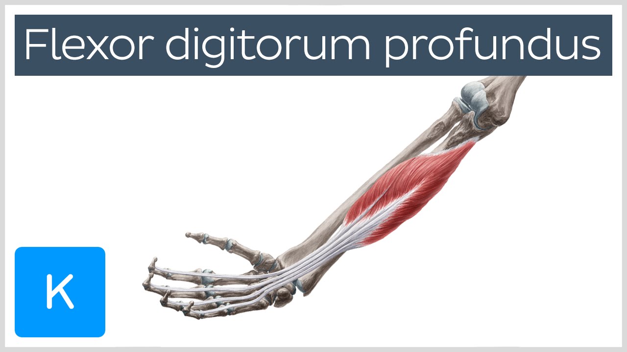
Flexor digitorum superficialis (fds) and flexor digitorum profundus (fdp). Both forearm bones articulate with the carpal bones of the wrist distally. In human anatomy, the flexor digitorum profundus (fdp, latin for deep bender of the fingers) is a muscle in the forearm that flexes the fingers (also known as digits). Hand and finger ultrasound education showing how to, scanning protocol, normal anatomy, anatomic variants, tendon, pulley googhywoiu9839t543j0s7543uw1. (ulnar half), abductor digiti minimi m., flexor digiti minimi brevis m., opponens digiti minimi m., ulnar 2 lumbrical mm., palmar and dorsal interosseous mm. It can be treated by simple fasciotomy. Quadrigia effect or dip joint flexion contracture occurs when profundus advancement of 1 cm or more. The muscle that moves these tendons is a common muscle belly shared by all the fingers. Quiz 2533 j hand surg am. Flexion of fingers is done by two tendons viz. Each finger has two flexor tendons, the flexor digitorum superficialis (fds) and the flexor digitorum profundus (fdp). The forearm consists of two bones, the radius and the ulna. These tendons enter the hand via the carpal tunnel, enclosed in a common synovial sheath.
This complex structure connects the entire hand to the radius and ulna, facilitates the passage of tendons together with the above mentioned neurovascular structures from the forearm to the hand, and permits us to exploit all its movements. The muscle that moves these tendons is a common muscle belly shared by all the fingers. This muscle extends from the proximal part of the ulna to the distal phalanges of the 2nd to 5th digit. There, the tendons are surrounded by several layers of loose connective tissue. Both forearm bones articulate with the carpal bones of the wrist distally.
Flexor Digitorum Profundus Muscle Origin Insertion Innervation Function Anatomy Kenhub Youtube from i.ytimg.com Kaplan's functional and surgical anatomy of the hand. Flexor digitorum superficialis (fds) and flexor digitorum profundus (fdp). Flexion of fingers is done by two tendons viz. The extensor indicis proprius and extensor digiti minimi lie ulnar to the extensor. Compartment syndrome rarely occurs in the first dorsal interosseous compartment of hand. Within the fingers, the fds tendons split at the level of the mid diaphysis of the proximal phalanges to allow the fdp tendons to pass superficially. The long flexor tendons of the fingers arise from the flexor digitorum superficialis (fds) and flexor digitorum profundus (fdp) forearm muscles. Ebraheim's educational animated video describes the anatomy of the flexor digitorum profundus muscle.the flexor digitorum profondus is a muscle in the fo.
Biology and anatomy of flexor tendon lecture.
Muscles flexor digitorum profundus (fdp) functions as a flexor of the dip joint; Quiz 2533 j hand surg am. Compartment syndrome rarely occurs in the first dorsal interosseous compartment of hand. Leddy and packer classification for zone i flexor tendon injury: Hand and finger ultrasound education showing how to, scanning protocol, normal anatomy, anatomic variants, tendon, pulley googhywoiu9839t543j0s7543uw1. Within the hand, the tendons fan out and enter their respective fibrous flexor sheaths. The long flexor tendons of the fingers arise from the flexor digitorum superficialis (fds) and flexor digitorum profundus (fdp) forearm muscles. It is considered an extrinsic hand muscle because it acts on the hand while its muscle belly is located in the forearm. Within the wrist and hand, the fds tendons are located superficially (volar) to the fdp tendons. The muscle that moves these tendons is a common muscle belly shared by all the fingers. Anatomy of the flexor tendon sheath and pulley system: The muscle belly divides into 4 tendons. This complex structure connects the entire hand to the radius and ulna, facilitates the passage of tendons together with the above mentioned neurovascular structures from the forearm to the hand, and permits us to exploit all its movements.
The forearm consists of two bones, the radius and the ulna fdp anatomy. It is the mass action muscle so act as the main gripping power of the hand because the tendons of the flexor digitorum profundus arise at or below the wrist joint (whereas tendons of flexor digitorum superficially arise in distal 3rd of the forearm).


Comments
Post a Comment